Diagram Of The Compound Light Microscope Microscope Diagram
Compound light microscope parts labeled The microscope (lesson 0362) [diagram] mercury labeled diagram
regardless of rupture digestion light microscope labeled diagram
Microscope light diagram parts drawing labeled compound biology sketch labelled microscopes cell worksheet google part textbook simple structure labeling search Microscope world blog: september 2015 Study of compound microscope
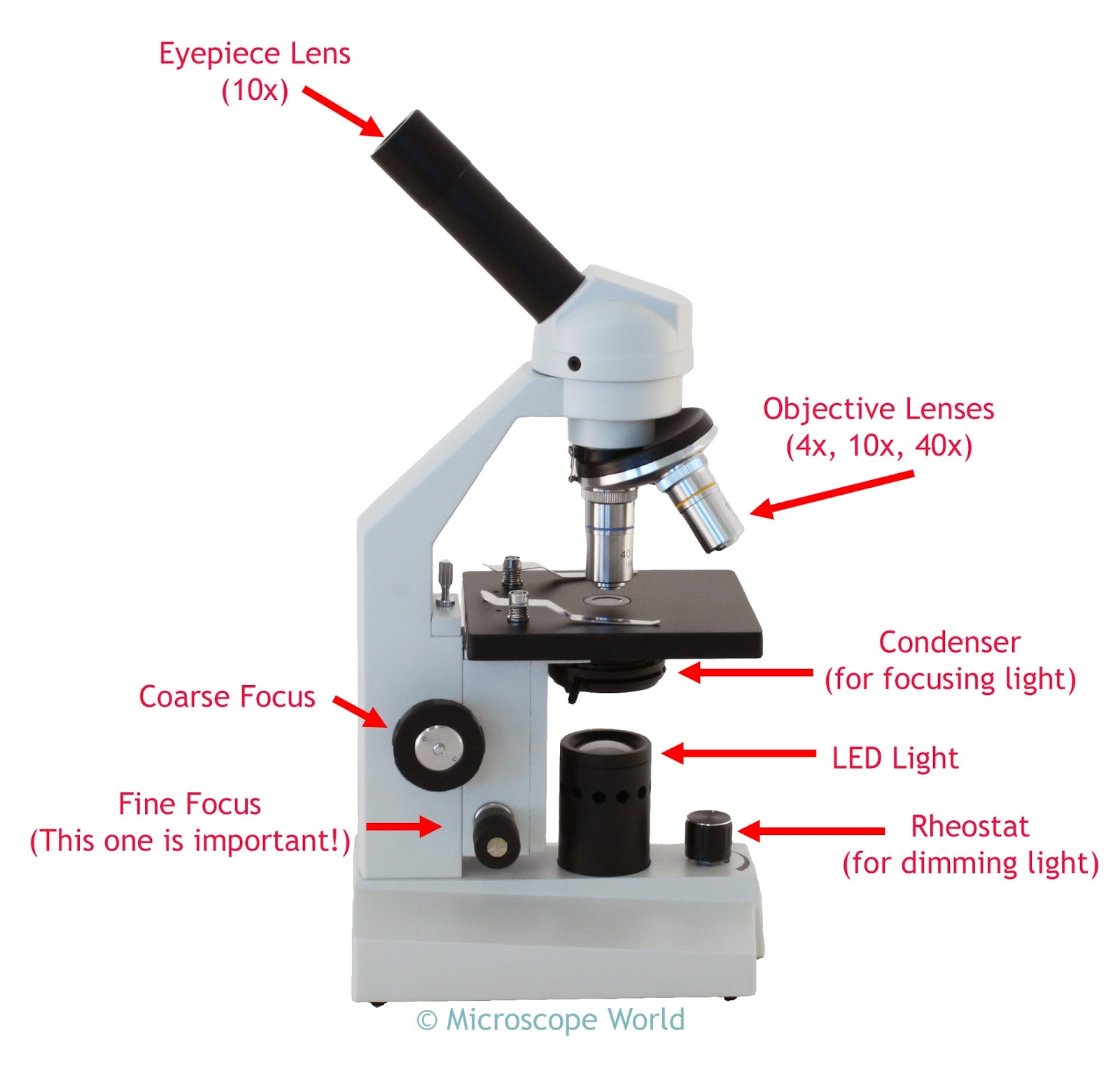
Parts of a compound light microscope rheostat
Compound light microscope functions of partsMicroscope simple parts optical microscopes uses principle actually drawing functions mechanical components definition supports several made Microscope labeled blank unlabeledWhat is compound microscope?.
Compound light microscope labeled diagramParts of a compound light microscope worksheet Microscope light diagram parts drawing labeled microscopes compound biology cell labelled sketch cells structure google simple microscopic cronodon cliparts scientificCompound microscope light source.

5 types of microscopes with definitions, principle, uses, labeled diagrams
Book club for teens 2018-19Draw a ray diagram of compound microscope Parts of a microscopeAddgene: using a light microscope protocol.
Microscope drawing template compound quiz labeled light lab cell drawings paintingvalleyMicroscope parts diagram drawing light labeled compound biology labelled sketch microscopes part simple structure google textbook cliparts cronodon search labeling Microscope diagramParts of a compound light microscope worksheet.

Simple microscope: definition, principle, parts, and uses » microscope club
Regardless of rupture digestion light microscope labeled diagramMikroskop optyczny/optical microscope – phobia — photonics and Which structure is best observed using a compound light microscopeCells and microscopes.
Microscope diagram labeled, unlabeled and blankCompound microscope: diagram, parts, working & magnification Microscope use labeled label diagram microscopeworld prepared slides answers kids setThe cell.


microscope diagram - Google Search | Microscopic, Animal cells

Study of Compound Microscope - Solution Parmacy

Parts Of A Compound Light Microscope Rheostat

Cells and Microscopes

regardless of rupture digestion light microscope labeled diagram

Compound Light Microscope Functions Of Parts | My XXX Hot Girl

Compound Light Microscope Labeled Diagram
![What is Compound Microscope? - Diagram, Function [updated]](https://i2.wp.com/www.tutoroot.com/blog/wp-content/uploads/2022/09/Compound-microscope-1-300x273.png)
What is Compound Microscope? - Diagram, Function [updated]
Microscope World Blog: September 2015